Function of Xylem
Xylem transports water in plants and help to support them.
Function of Phloem
Phloem transports assimilates (substances made by the plant) from the source (i.e. areas where they are produced, like leaves) to a sink (areas where they are used or stored, like the root or the flower).
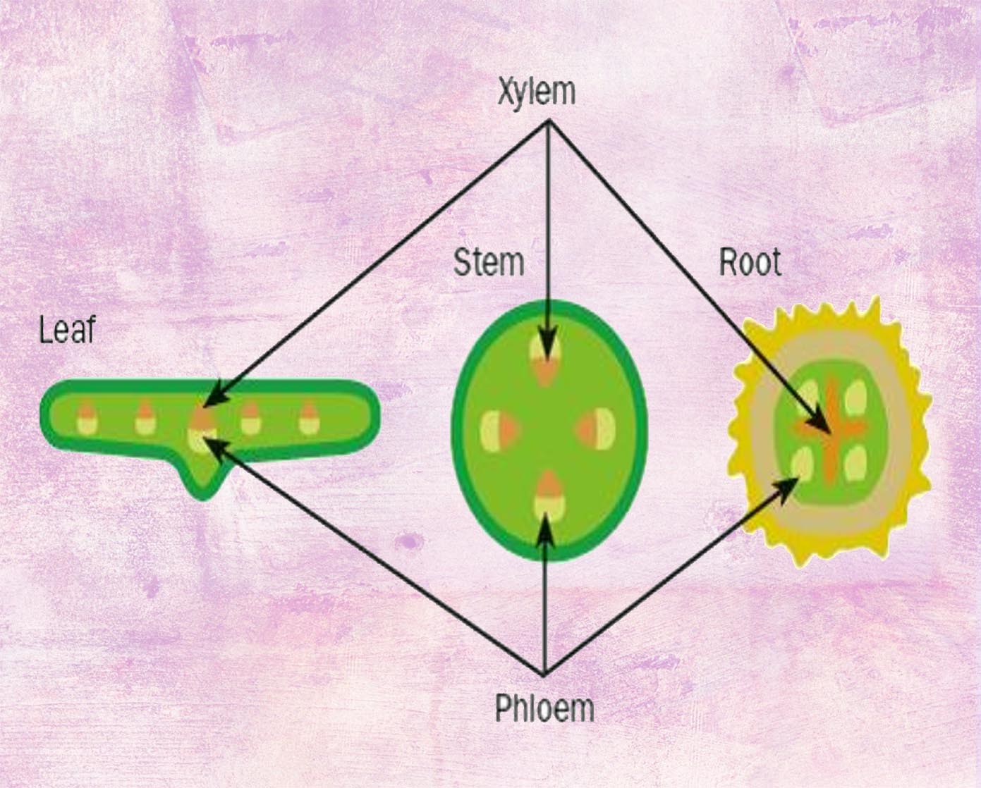
Position of xylem and Phloem in non-woody dicotyledonous plants
You are required to know the positions of xylem and phloem in leaf, stem as well as roots.
Root Hair Cells
Click on the root hair cell to find out its function.
[Supplement] Adaptation of root hair cells
The root hair in a root hair cells is essentially just a long finger-like projection from the cell. This increases the surface area of the cell. This helps speed up osmosis and the diffusion of mineral ions into the cell.
The large surface area of root hairs increases the rate of the absorption of water
Water Pathway
Water first diffuses into the root hair by osmosis, it then diffuses from cell to cell or cell wall to cell wall (or any other combination of cells and cell walls) through the root cortex, where it eventually reaches the xylem. Water is taken up through the xylem until it reaches a leaf, where it diffuses out into the surrounding mesophyll cells. Water diffuses from the mesophyll cells to the surrounding intercellular air spaces as water vapour, and finally, out of the leaf through the stomata.
Water Pathway Summary
The whole process can also be visualised using this diagram.
Experiment: Water Pathway
Cut the base of a stalk of celery (the non-leafy end) under water. Place the stalk in a beaker of water that has been stained with red food dye, the base end down, and place it in room temperature conditions, under a bright light and a slight breeze. You may observe red lines travelling up the stalk, and then through the leaves. If you cut the stalk halfway, you can also observe where the xylem vessels
Transpiration
Transpiration is loss of water vapour from plant leaves by evaporation of water at the surfaces of the mesophyll cells followed by diffusion of water vapour through the stomata.
Factors affecting Transpiration: Humidity
The diffusion of water vapour is dependent on its concentration gradient between the leaf and outside air. As humidity decreases, the concentration gradient between the leaf and outside air becomes greater, and rate of transpiration increases.
Factors affecting Transpiration: Temperature
Transpiration is faster in higher temperatures
[Supplementary] Explanation for Humidity
Diffusion of water vapour out of the leaf slows down if the leaf is already surrounded by moist air.
[Supplementary] Explanation for Temperature
Evaporation and diffusion are faster at higher temperatures because water vapour molecules will have more kinetic energy and hence move out of the leaf faster.
Definition: Circulatory System
The circulatory system is a system of tubes with a pump and valves to ensure one-way flow of blood. The tubes are our blood vessels (veins, arteries, etc.), the pump is the heart, and we have valves in our veins and heart to ensure that blood won
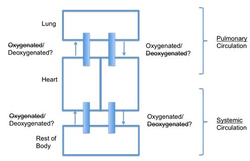
Double Circulation
Humans have a double circulation system. The first is the pulmonary circulation (towards the lungs) while the second is the systemic circulation(towards the rest of the body). The main function of our blood circulatory system is to transport nutrients and oxygen to all parts of the body and the removal of metabolic waste. Nutrients from digested food are transported from the intestines to other parts of the body while excretory metabolic waste are transported from all parts of the body to the kidney.
Double circulation means that the blood flows through two circuits
[Supplementary] Advantage of double circulatory system
The advantage of having a double circulation system is that blood in the two circulation can be at different pressures.
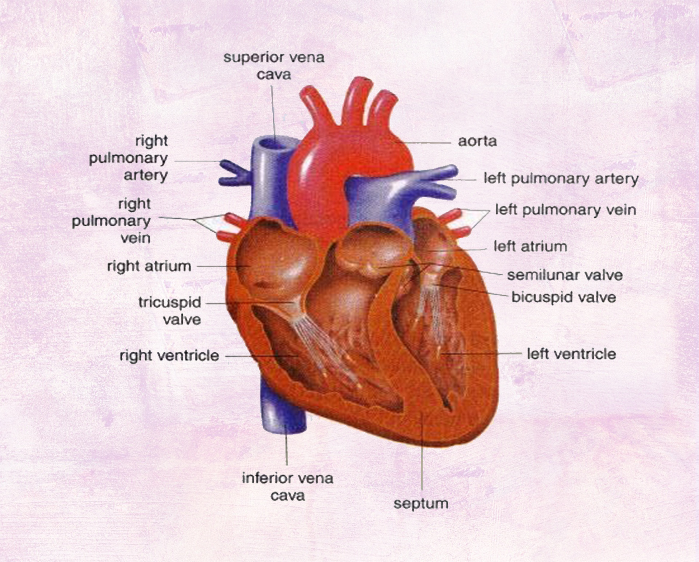
Heart Structure
You are required to name and identify the structures of the mammalian heart, limited to the muscular wall, the septum, the left and right ventricles and atria, one-way valves and coronary arteries. Note that all the diagrams here are not a mirror reflection of your own heart
Blood Flow
Blood is pumped away from the heart into arteries and returns to the heart in veins.
[Supplementary] Functioning of the heart
1. Blood from the body arrives at the heart via the vena cava, and enters the right atrium.
2. Contraction of the right atrium passes blood through the tricuspid valve to the right ventricle.
3. Contraction of the right ventricle forces blood out through the pulmonary artery to the lungs. The tricuspid valve closes to prevent backflow of blood into the right atrium, and the semi-lunar valve then closes to prevent the backflow of blood into the ventricle.
4. Blood enters the left atrium from the lungs theough the pulmonary vein.
5. Contraction of the left atrium passes blood to the left ventrivle through the bicuspid valve.
6. Contraction of the left ventricle forces blood out through the aorta towards the rest of the body. the bicuspid valve closes to prevent backflow of blood into the left atrium, amd then the semi-lunar valve closes to prevent backflow of blood into the ventricle.
Blood vessels to and from heart and lungs
The heart and the systemic circulation are connected by the aorta and vena cava while the heart and the pulmonary circulation are connected by the pulmonary artery and pulmonary vein.

[Supplementary] Coronary Heart Disease
Coronary heart disease (CHD) is caused by artherosclerosis. This is when plaque builds up in your arteries, thereby narrowing or blocking them up. This plaque is made of cholesterol, fatty substances, cellular waste products, calcium and fibrin. This buildup of plaque usually takes several years. Overtime, the plaque may harden, reducing the flow of oxygen rich blood to the heart muscles. Sometimes, this plaque might rupture (break apart), causing the formation of blood clots, or the broken piece of plaque to travel down to a narrow arteriole and block it up. Both of these completely cut off the supply of oxygen to the heart muscles. This could cause heart failure, which is diagnosed as CHD. CHD can be caused by a diet high in fat, especially saturated fats, it can be caused by stress, smoking, and sometimes, it is hereditary. In order to avoid CHD, it is important to maintain a balanced diet, have plenty of exercise, avoid unhealthy habits like smoking. Taking time to yourself and rel
Effect of physical activity on pulse rate
To investigate the effect of physical activity on pulse rate, we first have to know how to measure a pulse rate. This can be done by pressing two fingers down on the inside of their wrist, between the bone and the tendon on their thumb side. You should be able to feel pulses. This is because, during ventricular systole, blood is forced down the arteries, so they expand slightly. Using a stopwatch, count the number of pulses in 30 seconds, and multiply by two to gain the pulse rate per minute. Follow the steps as listed below to compare how physical activity affects pulse rate.
Step 1
Measure the pulse rate of a person at rest.
Step 2
Undergo exercise, e.g. jogging for 1 to 5 minutes.
Step 3
Measure their pulse rate again and compare.
[Supplementary] Explaination of effect of physical activity on pulse rate
Physical activity means the increased usage of muscles. As the muscles are working more, they require more energy, and hence, need more oxygen. This means that more blood needs to be pumped to the muscular tissue, so the heart works faster, hence increasing the pulse rate.
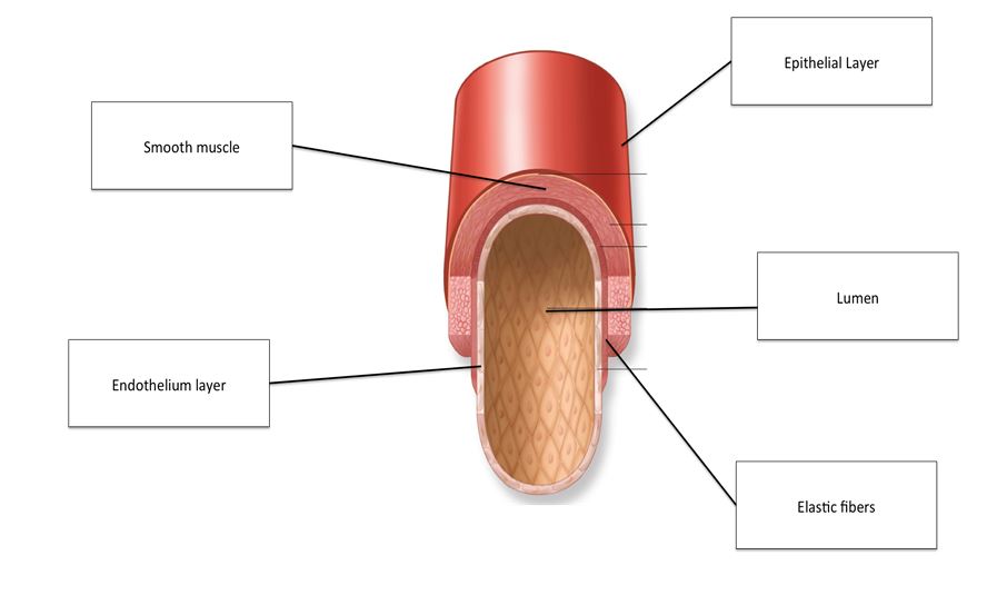
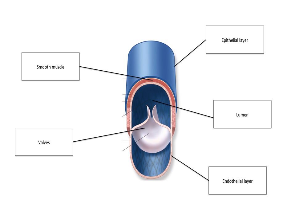
Arteries, veins and capillaries
All three blood vessels have a single layer of endothelial cells (squamous epithelium). Arteries and veins have a tunica media (smooth muscle) and a tunica externa (elastin and collagen). Arteries have more elastin than veins in their tunica externa. Arteries have a relatively small lumen (space inside the vessel), and veins have a larger lumen. Capillaries have the smallest lumen.
Artery
Arteries are blood vessels that transport blood away from the heart. Other than the pulmonary arteries, they carry oxygenated blood. They are thicker and more elastic compared to veins as they are exposed to higher pressure and force.
Arteries have the highest-pressure blood flowing through them, so they have the thickest walls.
Muscular contractions from the heart and the recoil from expansion and contraction of the artery drive the flow of blood in arteries. Compared to veins, their diameter is smaller, so blood pressure within the vessel is higher and flow faster. Thus, arteries have no valves.
Vein
Veins are blood vessels that transports blood to the heart. Other than the pulmonary veins, they carry deoxygenated blood. Veins have blood at a lower pressure flowing through them, so their walls don
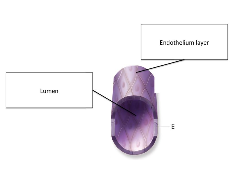
Capillary
Capillaries are very small in diameter, allowing them to bring blood closer to the needy tissues. This reduces the diffusion distance of oxygen from RBCs to tissue, and carbon dioxide from the tissue to the RBCs. They have a single cell thick wall, again reducing diffusion distance, and blood flows relatively slowly through arteries, allowing more time for diffusion.
Capillaries have a
Components of blood
Blood is made up of red blood cells, white blood cells, platelets and plasma.
Red Blood Cells & White Blood Cells
Click on the RBC and WBC to view how it looks under a light microscope.
Types of White Blood Cells
Function of Red Blood Cells
Red Blood Cells are made of many thousands of haemoglobin, each of which are made of four polypeptides. Each polypeptide has one iron ion (Fe2+) attached to it. This is where an oxygen molecules binds. This means, each haemoglobin molecule can carry up to 4 oxygen molecules and hence, 8 oxygen atoms.
When the RBCs are in the lungs, they are surrounded by a high concentration of oxygen, leading to more and more oxygen binding with the haemoglobin. This leads to the blood becoming saturated with oxygen
Function of White Blood Cells
Phagocytosis is the ingestion and digestion of bacteria by white blood cells. This successfully breaks down the pathogen into its harmless components, usually by neutrophils.
The stages of phagocytosis:
1.Ingestion: the bacteria/ food particles are engulfed by the WBC. This results in the formation of a food vacuole.
2. Vesicles, called lysosomes, containing digestive enzymes, fuse with the food vacuole, dumping the enzymes into said vacuole.
3. The bacteria are digested.
4. The components of the bacteria are often egested (dumped outside the WBC).
Antibodies are formed by lymphocytes.
The functions of antibodies include:
Act as a label: Cells with antibodies binded to them can be identified as target cells by phagocytes.
Neutralisation: the binding of an antibody to a pathogen can often cause the neutralisation of harmful toxins they release.
Agglutination: They may cause several pathogens to stick together, preventing them from dividing a
Function of Platelets
Platelets are small disc shaped cell fragments, that are involved in blood clotting.
Function of Plasma
About 55% of blood is plasma. It transports blood cells, ions, soluble nutrients, hormones and carbon dioxide. This is the solution that carries blood cells, and other solutes around the body. Plasma is a pale-yellow sticky liquid. It is 92% water, 8% dissolved protein, soluble nutrients, hormones and carbon dioxide. It takes RBCs close to respiring tissue to supply them with oxygen, it takes carbon dioxide from the respiring tissue and to the lungs, it carries nutrients from their sites of production to their sites of usage or storage, and transports hormones to their target organs.
